Ultrasound Tissue Characterisation
A 3D Scan and Powerful Tool for Achilles & Patellar Tendon Diagnosis & Management.
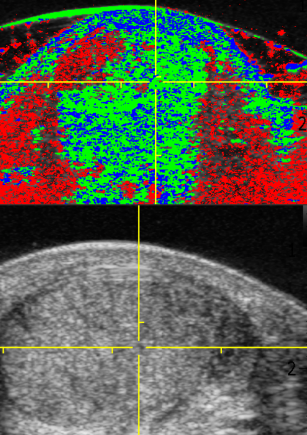
Tendon pain is a common complaint that can significantly impact an individual’s quality of life. Understanding the underlying cause of this pain is crucial for effective treatment. While physical examination and imaging techniques like ultrasound are valuable, Ultrasound Tissue Characterisation has emerged as a powerful diagnostic tool. This presentation will delve into the anatomy and function of the Achilles tendon, explore the causes of Achilles tendon pain, and discuss the role of ultrasound tissue characterisation in its diagnosis and informing management.
This clinic is only one of two centres in the UK offering this specialist tendon scan. I was trained by Professor D Hans Van Schie in the Netherlands and Jarrod Antflik in the UK.

Anatomy and Function of the Achilles Tendon
Anatomy
Function
Blood Supply
The Achilles tendon is the thickest and strongest tendon in the human body. It connects the calf muscles (gastrocnemius and soleus) to the heel bone (calcaneus). This tendon plays a crucial role in walking, running, and jumping by allowing plantar flexion of the foot.
During activities like walking or running, the Achilles tendon transmits the force generated by the calf muscles to the foot, enabling us to propel ourselves forward. A healthy Achilles tendon allows for smooth and efficient movement.
The Achilles tendon has a relatively poor blood supply, which means it takes longer to heal compared to other tissues. This limited blood flow makes the tendon prone to injury and degeneration.
Role of Ultrasound Tissue Characterisation (UTC)
Ultrasound tissue characterisation plays a crucial role in diagnosing and understanding the nature of Achilles tendon pain. It provides a non-invasive, readily available, and cost-effective way to visualize the tendon’s structure and identify abnormalities that may be causing pain. Due to its detail it helps guide the best treatment for your tendon.

UTC Scan is able to provide:
– Grey scale 3D Scan of the tendon
– UTC Analysis of tendon fibre types
– Statistical analysis of tendon fibres
– Accurate identifiction of affected fibres and how to target rehabilitation
Your content goes here. Edit or remove this text inline or in the module Content settings. You can also style every aspect of this content in the module Design settings and even apply custom CSS to this text in the module Advanced settings.
Clinical Applications
Diagnosis:
UTC helps differentiate between different causes of Achilles tendon pain, guiding appropriate treatment decisions.
Treatment monitoring:
UTC can monitor the healing process of the tendon, track the effectiveness of treatment, and identify any potential complications.
Pre & Post Operative Evaluation:
UTC is often used to assess the severity of tendon injuries before surgery, helping surgeons plan the appropriate surgical approach.
Training Programme Evaluation:
UTC is used to evaluate the tendon during and after specific training programmes ensuring optimal training for your tendon health.
Achilles Tendon or Patellar Tendon : Scan & Analysis
- Ultrasound Tissue Characterisation Single* Tendon Scan
- Ultrasound Tissue Characterisation Report with Images
Price £300
Elite Sport Package £550
* To scan both left and right tendons the fee is £500
Please note all UTC Scan must be paid for in advance. Please check with your insurance provider if scans are covered. Thank you.
About
Gafin Morgan, Consultant Podiatrist, Chartered Scientist (The Science Council), MS Sonographer (CASE Acc), Member Royal College of Podiatry, HCPC
Research Interests
Achilles tendinopathy, Tendon trauma healing, Elastography, Ultrasound Tissue Characterisation